Visualizing Seaweed Cell Walls with Bioorthogonal Chemistry
December 18, 2025
A groundbreaking study by Delpont, Neutelings, and Popper, recently published in Annals of Botany (Volume 136, Issue 2, July 2025), pioneers a powerful solution to this problem. Their work, aptly titled "Click & sea: using bioorthogonal click chemistry to visualize seaweed cell walls," successfully adapts and applies strain-promoted alkyne–azide cycloaddition (SPAAC), a type of bioorthogonal click chemistry, to visualize polysaccharide incorporation in living seaweed tissues for the first time. This methodological breakthrough opens new horizons for phycology, enabling real-time observation of cell wall dynamics in response to environmental stimuli, developmental cues, and the stresses of climate change.
Introduction
Seaweeds, the diverse and abundant photosynthetic organisms of our coastal ecosystems, represent an immense and largely untapped reservoir of unique biomolecules. Their cell walls, in particular, are a rich source of complex polysaccharides—such as ulvans in green seaweeds and carrageenans/agars in red seaweeds—with significant commercial value in food, pharmaceuticals, cosmetics, and biotechnology. However, studying the dynamic synthesis, architecture, and remodeling of these complex polymeric networks in situ has long posed a formidable challenge for researchers. Traditional methods, including histological staining and immunolabeling, are constrained by the limited availability of specific monoclonal antibodies against seaweed polysaccharide epitopes and the sheer structural diversity of these molecules.
The Critical Challenge: Studying Seaweed Cell Walls In Situ
Seaweed cell walls are not merely static structural supports; they are dynamic interfaces involved in growth, defense, and environmental interaction. Understanding their composition and metabolism is crucial, not only for fundamental biology but also for optimizing the extraction and application of their valuable polysaccharides. The commercial seaweed product market was valued at $5.3 billion in 2023 and is projected to grow significantly, underscoring the economic importance of these organisms.
Despite this, research tools have lagged behind. As detailed in the paper, the toolkit for in situ study is limited:
- Histological stains (e.g., Alcian Blue, Calcofluor White) provide general visualization but lack specificity for particular polysaccharide types or their metabolic states.
- Monoclonal antibodies offer high specificity, but their development for seaweed-specific components has been slow. Only a handful are commercially available (e.g., against alginates and fucoidans in brown seaweeds via SeaProbes), and many historically useful antibodies are no longer accessible. This is especially problematic for red seaweeds, whose commercially important carrageenans are poorly characterized at the cellular level.
This gap in methodology has hindered progress in understanding how seaweeds build and modify their cell walls throughout their life cycle or under stress conditions such as warming oceans, acidification, or pathogen attack.
The "Click & Sea" Methodology: A Step-by-Step Breakdown
Delpont and colleagues elegantly circumvented these limitations by leveraging metabolic labeling and bioorthogonal chemistry. Their approach is summarized in the following schematic from their publication:
- Selection of Biological Models and Reporters
The researchers chose two species with thin tissues to facilitate reagent uptake:
- Green seaweed: Ulva spp. (Sea Lettuce), known for its ulvan-rich cell walls and sequenced genome.
- Red seaweed: Phycodrys rubens, a species with a flattened thallus, chosen for its structural distinction.
They selected three azide-modified monosaccharide reporters—analogues of fucose, galactose, and glucose—based on their prominence as building blocks in major seaweed polysaccharides.
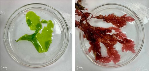 Fig.1 Specimens of the studied seaweed species, Ulva spp. (left) and Phycodrys rubens(right). (Delpont, et al., 2025)
- Metabolic Incorporation and Click Reaction
Small sections of seaweed were incubated in seawater containing these "reporter" sugars. The peracetylated form of the sugars allows them to passively diffuse into cells. Once inside, cellular metabolic enzymes process them, and they are incorporated into newly synthesized polysaccharides and glycoconjugates, introducing the azide "chemical handle" into the seaweed's biomolecules.
After an incubation period (24 or 48 hours), the unincorporated reporters were washed away. The seaweed tissues were then treated with a dibenzocyclooctyne (DBCO) reagent conjugated to the green-fluorescent dye. The DBCO group undergoes a rapid, specific, and copper-free SPAAC "click" reaction with the azide groups embedded in the cell walls, covalently attaching the bright fluorescent tag to the newly synthesized polymers.
- Visualization and Analysis
The labeled tissues were imaged using confocal microscopy. The resulting images revealed clear, specific fluorescence patterns.
Key Findings and Implications
The study yielded several important results:
Both Ulva and Phycodrys effectively incorporated the three sugar reporters, with fluorescence intensity increasing over a 48-hour period, indicating active metabolic uptake.
Fluorescence was predominantly associated with the cell walls, confirming that the reporters were integrated primarily into structural polysaccharides rather than just energy storage molecules. Intracellular fluorescence likely corresponds to organelles involved in polysaccharide synthesis (e.g., Golgi apparatus) or storage granules (e.g., starch).
- Species-Specific Patterns
In Ulva, glucose incorporation was the most intense, consistent with its role in cellulose and ulvan biosynthesis. In Phycodrys, all reporters showed strong incorporation, with intracellular patterns suggesting active synthesis of cell wall components like carrageenans (galactose-rich).
This work proves that bioorthogonal click chemistry is a viable, powerful, and relatively simple technique for studying seaweed cell walls. It bypasses the need for specific antibodies and allows for pulse-chase experiments to track polysaccharide deposition over time.
This technique can now be applied to answer critical questions: How do green tides form so rapidly? How does ocean acidification affect the structural integrity of seaweed cell walls? How do seaweeds remodel their walls during development or in response to grazing?
Bridging Research and Application: The GlycoCLICK™ Advantage
The "Click & Sea" study is a testament to the transformative power of click chemistry in glycobiology. While the researchers synthesized their own reagents, implementing such sophisticated techniques can be a significant hurdle for many labs. This is where specialized service providers can accelerate discovery.
GlycoCLICK™, a dedicated platform from CD BioGlyco, offers a comprehensive suite of services that directly build upon the principles demonstrated in this pioneering work. We provide end-to-end support for researchers looking to apply click chemistry to their own projects in marine biology and beyond:
We provide custom synthesis of azide- or alkyne-functionalized monosaccharide reporters, like the fucose, galactose, and glucose analogues used in the study, ensuring high purity and reliability.
Our experts can assist with experimental design for metabolic labeling of cells and tissues, including optimizing incubation times and conditions, just as was critical for the success with Ulva and Phycodrys.
We offer services for the subsequent click reaction with fluorescent probes and high-resolution confocal microscopy, delivering clear, analyzable images of glycan localization.
For applied research, we can help functionalize surfaces or nanoparticles with seaweed polysaccharides using click chemistry, facilitating the development of new drug delivery systems or bioactive materials.
By leveraging GlycoCLICK™ services, researchers can overcome technical barriers and focus on their biological questions, whether they involve fundamental seaweed physiology or the development of the next generation of seaweed-based products.
Conclusion
The "Click & Sea" methodology marks a significant leap forward for marine science. It provides phycologists with a versatile and specific tool to visualize and understand the dynamic world of seaweed cell walls. As we face the challenges of climate change and seek sustainable bio-resources from the ocean, such innovative techniques are invaluable.
The fusion of basic research, as showcased in this seminal paper, with advanced technological platforms like GlycoCLICK™, paves the way for a deeper understanding of glycans and their profound role in nature and industry.
Reference
- Delpont, W.F.; et al. Click & sea: using bioorthogonal click chemistry to visualize seaweed cell walls. Annals of Botany. 2025, 136(2): 437-450.